Sat 27 January 2024:
In a study published in Science Advances on Jan. 24, researchers led by Prof. ZHU Maoyan from the Nanjing Institute of Geology and Palaeontology of the Chinese Academy of Sciences reported their recent discovery of 1.63-billion-year-old multicellular fossils from North China.
These exquisitely preserved microfossils are currently considered the oldest record of multicellular eukaryotes. This study is another breakthrough after the researchers’ earlier discovery of decimeter-sized eukaryotic fossils in the Yanshan area of North China, and pushes back the emergence of multicellularity in eukaryotes by about 70 million years.

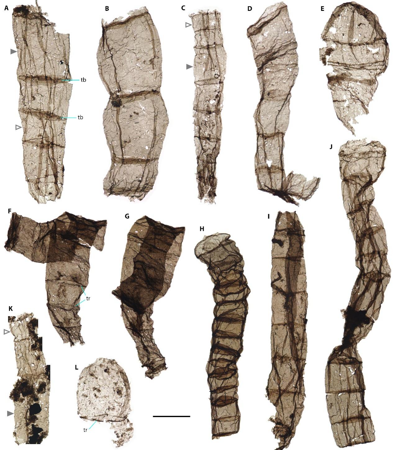
Multicellular fossil Qingshania from the Chuanlinggou Formation. Scale bar equals 100 μm for F-H and K, and 50 μm for the rest. (Image by MIAO et al.)
All complex life on Earth, including diverse animals, land plants, macroscopic fungi, and seaweeds, are multicellular eukaryotes. Multicellularity is key to eukaryotes acquiring organismal complexity and large size, and is often regarded as a major transition in the history of life on Earth. However, scientists have been unsure when eukaryotes evolved this innovation.
Fossil records offering convincing evidence show that eukaryotes with simple multicellularity, such as red and green algae, and putative fungi, appeared as early as 1.05 billion years ago. Older records have claimed to be multicellular eukaryotes, but most of them are controversial because of their simple morphology and lack of cellular structure.
“The newly discovered multicellular fossils come from the late Paleoproterozoic Chuanlinggou Formation that is about 1,635 million years old. They are unbranched, uniseriate filaments composed of two to more than 20 large cylindrical or barrel-shaped cells with diameters of 20–194 μm and incomplete lengths up to 860 μm. These filaments show a certain degree of complexity based on their morphological variation,” said MIAO Lanyun, one of the researchers.
The filaments are constant, or tapered throughout their length, or tapered only at one end. Morphometric analyses demonstrate their morphological continuity, suggesting that they represent a single biological species rather than discrete species. The fossils have been named Qingshania magnifica, 1989, a form taxon with similar morphology and size, and are described as being from the Chuanlinggou Formation.

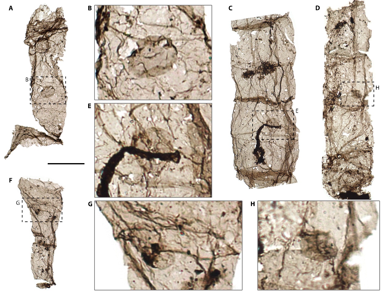
Multicellular fossil Qingshania with a round intracellular structure interpreted to be spore. Scale bar equals 50 μm for A, C, D and F. (Image by MIAO et al.)
A particularly important feature of Qingshania is the round intracellular structure (diameter 15–20 μm) in some cells. These structures are comparable to the asexual spores known in many eukaryotic algae, indicating that Qingshania probably reproduced by spores.
In modern life, uniseriate filaments are common in both prokaryotes (bacteria and archaea) and eukaryotes. The combination of large cell size, wide range of filament diameter, morphological variation, and intracellular spores demonstrate the eukaryotic affinity of Qingshania, as no known prokaryotes are so complex. Filamentous prokaryotes are generally very small, about 1–3 μm in diameter, and are distributed across more than 147 genera of 12 phyla. Some cyanobacteria and sulfur bacteria can reach large sizes, up to 200 μm thick, but these large prokaryotes are very simple in morphology, with disc-shaped cells, and are not reproduced by spores.
The best modern analogues are some green algae, although filaments also occur in other groups of eukaryotic algae (e.g., red algae, brown algae, yellow algae, charophytes, etc.), as well as in fungi and oomycetes.
“This indicates that Qingshania was most likely photosynthetic algae, probably belonging to the extinct stem group of Archaeplastids (a major group consisting of red algae, green algae and land plants, as well as glaucophytes), although its exact affinity is still unclear,” said MIAO.
In addition, the researchers conducted Raman spectroscopic investigation to test the eukaryotic affinity of Qingshania from the perspective of chemical composition, using three cyanobacterial taxa for comparison. Raman spectra revealed two broad peaks characteristic of disordered carbonaceous matter. Furthermore, the estimated burial temperatures using Raman parameters ranged from 205–250 °C, indicating a low degree of metamorphism. Principal component analysis of the Raman spectra sorted Qingshania and the cyanobacterial taxa into two distinct clusters, indicating that carbonaceous matter of Qingshania is different from that of cyanobacterial fossils, further supporting the eukaryotic affinity of Qingshania.
Currently, the oldest unambiguous eukaryotic fossils are unicellular forms from late Paleoproterozoic sediments (~1.65 billion years ago) in Northern China and Northern Australia. Qingshania appeared only slightly later than these unicellular forms, indicating that eukaryotes acquired simple multicellularity very early in their evolutionary history.
Since eukaryotic algae (Archaeplastids) arose after the last eukaryotic common ancestor (LECA), the discovery of Qingshania, if truly algal in nature, further supports the early appearance of LECA in the late Paleoproterozoic—which is consistent with many molecular clock studies—rather than in the late Mesoproterozoic of about 1 billion years ago.
This study was funded by the National Key Research and Development Program of China, the National Natural Science Foundation of China, and the Innovation Cross-Team of CAS.

Simplified eukaryotic tree (A) and representative early eukaryotic fossils (B). In eukaryotic tree, grey dash lines represent stem group eukaryotes. Solid lines denote crown group eukaryotes (LECA plus its descendants). Grey bars at nodes display the estimated age range of divergence of major branches from a molecular clock study (Parfrey et al.). Scale bar in the green algal fossil equals 500 μm; the rest are 50 μm. (Image by MIAO et al.)
Source: Chinese Academy of Sciences
______________________________________________________________
FOLLOW INDEPENDENT PRESS:
WhatsApp CHANNEL
https://whatsapp.com/channel/0029VaAtNxX8fewmiFmN7N22
![]()
TWITTER (CLICK HERE)
https://twitter.com/IpIndependent
FACEBOOK (CLICK HERE)
https://web.facebook.com/ipindependent
Think your friends would be interested? Share this story!


