
Thu 05 August 2021:
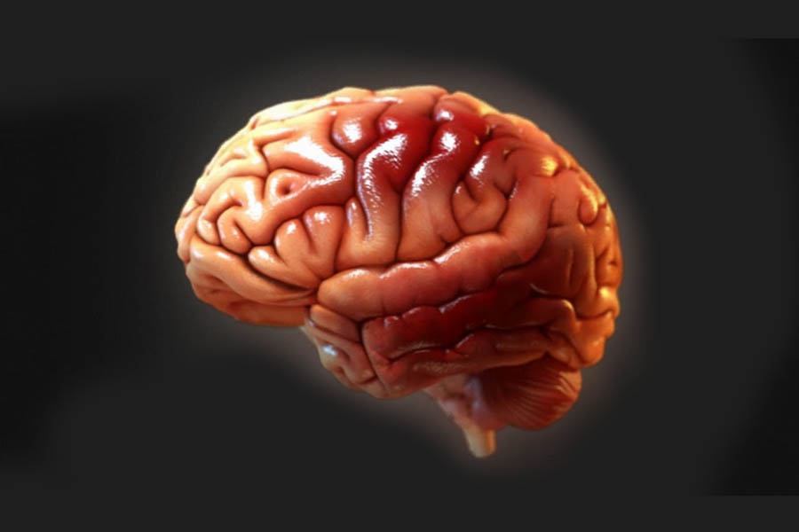
Scientists have unveiled the world’s first high-resolution 3D image of a monkey brain, which could open the door for treatments for human disorders including Parkinson’s disease.
A team from the Chinese Academy of Sciences in Beijing used fluorescence imaging techniques to generate a precise map of a full macaque monkey brain. According to The Daily Mail, “the team employed a new approach to reveal how nerve cells are organized and connected within the monkey brain at a micron resolution.”


The human brain comprises nearly a hundred billion nerve cells with delicate and complex connections, and while up to 17 times larger than that of a macaque, it is similar enough for comparisons to be made between the two, researchers claim.
Until now, a mouse brain was the largest to be mapped, taking days to create a complete 3D image, but the new technique made it possible to move up to a macaque brain, which is about 200 times larger in volume than that of a mouse. The team, including researchers from Zhejiang University, says that having such a detailed map of a primate brain will help in understanding human diseases.
The new imaging technique allowed the team to map every neuron and fiber of the monkey brain in greater detail than previously possible. They were able to peer down to the single micron level, viewing brain cells typically 100 microns across in levels of detail never before observed in a primate brain. The resulting images are massive, taking up more than a petabyte of data – 1,000 terabytes or about 30million high-definition movies.
With billions of neurons captured in unprecedented detail, the team turned to artificial intelligence to study the results. To capture this detail they created a new technique, known as Volumetric Imaging with Synchronous on-the-fly-scan and Readout (VISoR).
Compared with commonly used 3D optical imaging techniques, VISoR eliminates the time loss caused by moving and pausing while switching fields of view.
This means that they can complete a 3D image of a much larger brain than was previously possible, the team explained. They can take a full image of a monkey brain in under four days – about the same time it previously took to capture a full mouse brain, which is 200 times smaller.
———————————————————————————————————————-
FOLLOW INDEPENDENT PRESS:
TWITTER (CLICK HERE)
https://twitter.com/IpIndependent
![]()
FACEBOOK (CLICK HERE)
https://web.facebook.com/ipindependent
![]()
Think your friends would be interested? Share this story!